Vascular
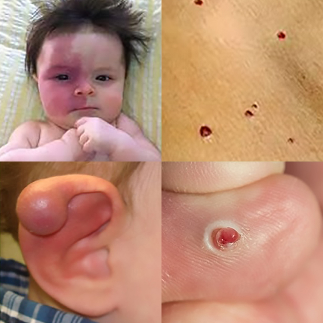
Vascular lesions are common benign lesions of the skin that can look alarming due to is colour. There are several different types of vascular lesion which vary in appearance (red or deep purple in colour) and treatment of the lesions depends on the origin of the lesion.
Types of vascular lesions:
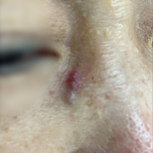
Hemangiomas are the most common type of vascular lesions in children. They are noncancerous and typically appears shortly after birth. They can progress through several phases: the growth phase, the resting phase, and the involution phase. Depending on how close these vascular lesions are to the surface of the skin, lesions can appear bright red (superficial hemangioma) to dark bruise like colour (deep hemangiomas). Superficial hemangiomas can grow towards the surface of the skin, causing ulceration and bleeding/infections.

Hemangiomas are the most common type of vascular lesions in children. They are noncancerous and typically appears shortly after birth. They can progress through several phases: the growth phase, the resting phase, and the involution phase. Depending on how close these vascular lesions are to the surface of the skin, lesions can appear bright red (superficial hemangioma) to dark bruise like colour (deep hemangiomas). Superficial hemangiomas can grow towards the surface of the skin, causing ulceration and bleeding/infections.
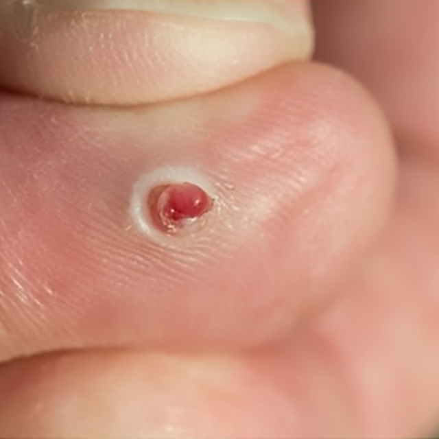
Hemangiomas are the most common type of vascular lesions in children. They are noncancerous and typically appears shortly after birth. They can progress through several phases: the growth phase, the resting phase, and the involution phase. Depending on how close these vascular lesions are to the surface of the skin, lesions can appear bright red (superficial hemangioma) to dark bruise like colour (deep hemangiomas). Superficial hemangiomas can grow towards the surface of the skin, causing ulceration and bleeding/infections.
Hemangiomas are the most common type of vascular lesions in children. They are noncancerous and typically appears shortly after birth. They can progress through several phases: the growth phase, the resting phase, and the involution phase. Depending on how close these vascular lesions are to the surface of the skin, lesions can appear bright red (superficial hemangioma) to dark bruise like colour (deep hemangiomas). Superficial hemangiomas can grow towards the surface of the skin, causing ulceration and bleeding/infections.
Vascular Malformations – a group of congenital errors where vessels were formed incorrectly. They are classified based on the type of vessels that make up the lesions.
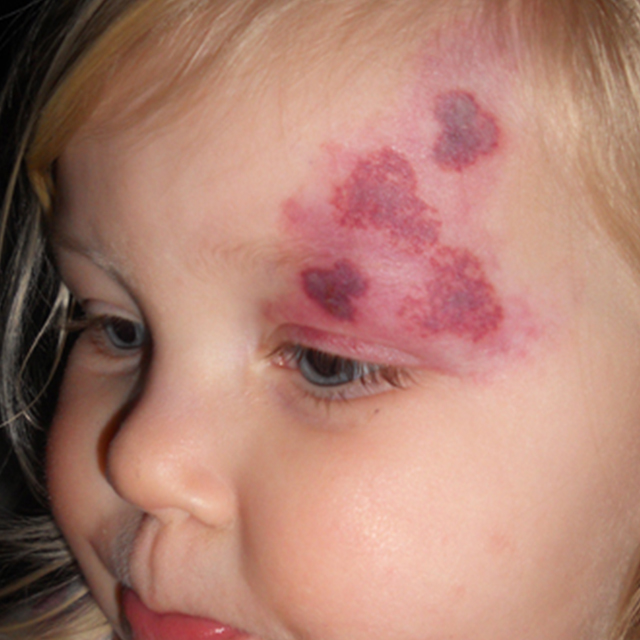
Capillary Malformations (also known as port wine stains) – made of irregular capillaries and small veins that run in the deeper areas of the skin. They appear as flat pick areas that can darken with time to deep purple in colour giving a cobble-stone like texture in the skin.
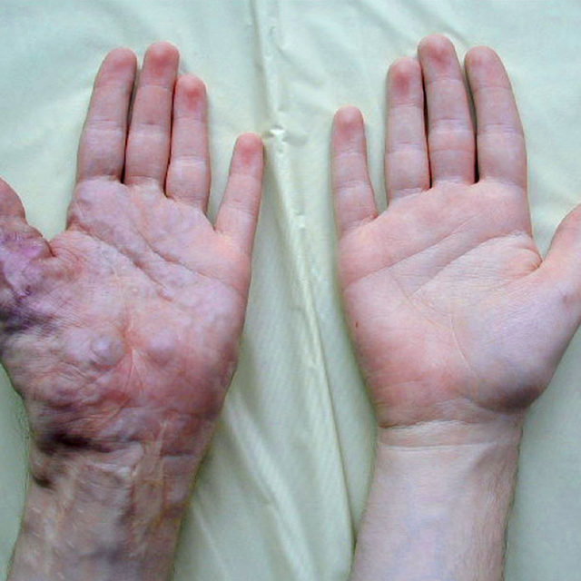
Venous Malformations – found in 1-4% of the population that is made of an abnormal collection of venous channels. They are bluish in appearance and can cause pain if clots form in the channels causing swelling in the area.
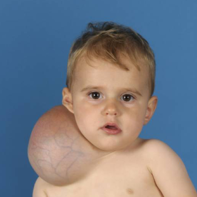
Lymphatic Malformation – made of an abnormal collection of lymphatic channels and vary in the range of appearance from small clear vesicles to cyst-like changes that are compressible and causes a bluish skin discolouration. If they are superficial, they can become infected causing discomfort and can cause disfigurement when they grow rapidly in size.
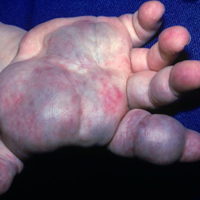
Arteriovenous Malformations – made of abnormal vascular connections between arteries and veins. They are high pressure systems where there is fast movement of blood through the systems. These channels can increase in time, becoming warm pulsating masses, and can cause significant problems such as increase demand on the heart, hemorrhage, and necrosis of surrounding tissues due to insufficient nutrient delivery to these tissues.
Vascular Malformations – a group of congenital errors where vessels were formed incorrectly. They are classified based on the type of vessels that make up the lesions.

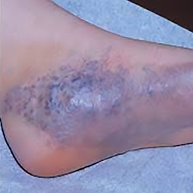

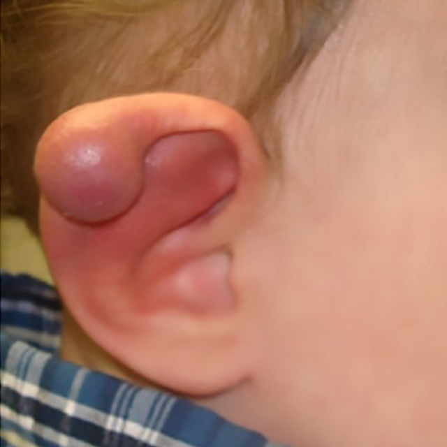

Pyogenic Granulomas – they typically require surgical management to completely remove the malformed vascular, either through excision or cauterization of the vessels, right to the base of the lesion, preventing the recurrence of the lesion.

Pyogenic Granulomas – they typically require surgical management to completely remove the malformed vascular, either through excision or cauterization of the vessels, right to the base of the lesion, preventing the recurrence of the lesion.

Hemangiomas: some studies have correlated hemangiomas with:
- Females at birth
- White, non-Hispanic ethnicity
- Premature birth
- History of chorionic villus sampling
Vascular Malformations: are typically congenital lesions (lesions from birth) and risk factors are largely unknown. In some rare instances, they are sometimes associated with genetic disorders, such Port-Wine Stains is often associated with Sturge-Weber disease.
Pyogenic Granuloma: typically associated with previous damage/trauma to the skin, chronic irritation to a certain area, capillary malformations, and laser treatment to capillary malformations.

Hemangiomas: there are several different approaches to management of hemangiomas depending on the life cycle of the lesion.
1. Medication – oral medications such as steroids of blood pressure medications can slow the growth of these lesions
2. Surgery – careful evaluation to await proper timing of surgical excisions must be performed. As some lesion can decrease in size, thus delay of surgical excisions, which can lead to scarring, is sometimes appropriate.
3. Laser Therapy – this type of therapy can be used to treat ulcerated lesions or residual lesions that remains after the lesions has progressed through the different phases.
Vascular Malformations: treatment is depending on the origin of the vascular malformation
1. Capillary Malformations: typically treated with serial laser treatments which targets the residual hemoglobin found in tissue gradually decreasing the discolouration found in the skin
2. Venous Malformations:
- Sclerotherapy – involves the injection of chemicals into the expanded venous tissue to cause collapse of the venous channel
- Surgical excision – is a direct removal and debulking of the malformation
3. Lymphatic Malformations
- Laser Therapy – typically used to treat small lesions as lasers tan seal off small oozing vessels
- Sclerotherapy – used to treat large lesions that maybe close to vital structures typically by interventional radiologist who can isolated troublesome vessels and target them to be collapsed by the chemicals injected into the vessel
- Surgical Excision – used to debulk large lesions often in combination with sclerotherapy
4. Arteriovenous Malformations: treatment of these lesions is often complex as these are high flow vessels. Treatment includes embolization (physically blocking) of the feeding vessels by interventional radiologists or vascular surgeons and/or complete excision of the lesion to prevent further recruitment of blood flow from other sources that may develop after embolization has occurred. Careful planning to explore different treatment approach is often needed for these types of lesions.
Pyogenic Granulomas: they typically require surgical management to completely remove the malformed vascular, either through excision or cauterization of the vessels, right to the base of the lesion, preventing the recurrence of the lesion.


