Melanocytic Naevi

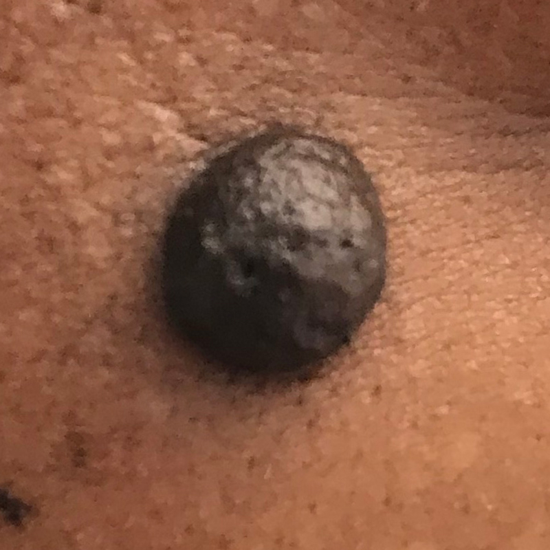
Melanocytic naevi are pigmented moles. “Melanocytic” means that they are made up of melanocytes, cells that produce the dark pigment, melanin, that gives skin its color. Melanocytes clustered together to form naevi. Melanocytic naevi can be found anywhere on the skin. Melanocytic naevi that are present at birth or developed within the first two years are called congenital melanocytic naevi. Giant congenital naevi that are larger than 20 cm in diameter have a greater risk for developing melanoma, which is cancerous. Melanocytic naevi that develop during childhood and early adult life are called acquired melanocytic naevi. Dysplastic or atypical naevi is less common and have an increased chance to develop into melanoma.
Risk
Signs and Symptoms
Diagnosis
Treatment
Management

- Genetic cause is suspected in congenital melanocytic naevi
- Sun exposure can increase the number of moles
- Possibly hormones or other biological processes may play a role in triggering acquired melanocytic nevi

- Genetic cause is suspected in congenital melanocytic naevi
- Sun exposure can increase the number of moles
- Possibly hormones or other biological processes may play a role in triggering acquired melanocytic nevi

- Acquired melanocytic naevi can be flat, circular, mild to dark brown with well defined borders; or raised, brown bumps and hairy with a slightly warty surface
- Moles can develop anywhere on the body including scalp, armpits, under the nails, between the fingers and toes.
Most people have 10 to 40 moles, usually asymptomatic and benign - Hormonal changes of adolescence and pregnancy may cause moles to become larger and darker
- Moles are usually less than 6 mm in diameter. Rarely, congenital nevi can be over 20 cm in diameter are called giant congenital melanocytic nevi. In rare cases melanoma may develop in this type of giant mole
- Atypical or dysplastic nevi are not common and have darker center and lighter, irregular borders and > 6mm in diameter. Although benign, they exhibit some clinical and histologic features of malignant melanoma so warrants close surveillance
- Person having 5 or more atypical naevi, or more than 50 melanocytic naevi have 6 times higher risk to develop melanoma

- Melanocytic Naevi are benign so can be left alone
- If cannot distinguish between atypical naevus and a melanoma, use of a dermoscopy would be helpful (using a dermatoscope, a hand-held high quality magnifying lens and a powerful lighting system to examine the skin surface)
- If in doubt, a suspicious or changing atypical naevus should be removed by excision biopsy and samples sent to the lab for histology assessment. Partial biopsy should be avoided

- Majority of melanocytic naevi do not need treatment due to its benign nature
- For giant congenital naevus, option of surgical removal if it causes emotional distress to the child or worry about risk of developing into melanoma
- Other option of mole removal would be excision, freeze with liquid nitrogen and laser treatment

- Closely monitor the atypical or giant congenital melanocytic naevi for changes in size and appearance
- Self- check of skin condition and visit family doctor or dermatologist for a thorough skin check when needed

- Closely monitor the atypical or giant congenital melanocytic naevi for changes in size and appearance
- Self- check of skin condition and visit family doctor or dermatologist for a thorough skin check when needed