Basal Cell Carcinoma (BCC)
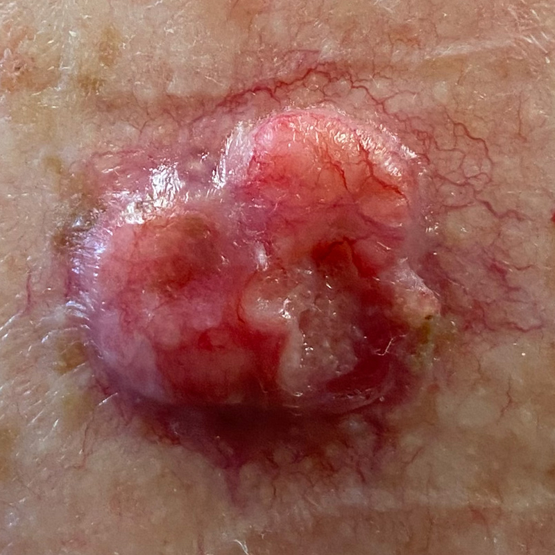
Basal cell carcinoma (BCC) is the most common type of skin cancer and makes up 75% to 80% of all skin cancers. Basel cells are found in the lower part of the skin and constantly divide to form new cells, replacing the older skin cells that sheds off the skin surface. Basal cell carcinomas often appear as slightly transparent lumps on the skin.
Risk
Prevention
Signs and Symptoms
Diagnosis
Treatment
Management

- UV light: Overexposure to ultraviolet radiation from sunlight or tanning beds.
- Genetics: Family history of skin cancer.
- Fair Skin: Having fairer skin reduces your protection from UV light.
- Weak immune system: Illnesses and immunosuppressive medications can weaken your immune system and prevent it from fighting off the SCC.
- Age/Sex: Being older will increase your chance of developing SCC, and men develop SCC at a higher rate than women.

- Avoid prolonged exposure under sunlight between 10 a.m. to 4 p.m., wear sunscreen at least SPF 50
- Wear protective clothing covering the arms and legs, broad-brimmed hat.
- Avoid tanning beds.
- Examine your skin often.
- Occurs most often on head and neck, less often on genitals.

- Avoid prolonged exposure under sunlight between 10 a.m. to 4 p.m., wear sunscreen at least SPF 50
- Wear protective clothing covering the arms and legs, broad-brimmed hat.
- Avoid tanning beds.
- Examine your skin often.
- Occurs most often on head and neck, less often on genitals.

Develop sores and non-healing growth on skin that appears:
- Pearly white, skin-colored, or pink bump that is translucent. Can appear on the face and ears. The lesion may breakdown, bleed or scab over.
- Brown, black, or blue lesion or with dark spots with a slightly raised, translucent border.
- Flat, scaly reddish patch with a raised edge, more common on the chest or back.
- White, waxy, scar-like lesion without a well-defined border, called morpheaform basal cell carcinoma.

- Doctor examines the suspicious skin lesion and the rest of the body.
- Skin biopsy, removing the lesion and review under the microscope.
- If biopsy results confirm BCC, examinations are ordered to rule out metastases.

- Doctor examines the suspicious skin lesion and the rest of the body.
- Skin biopsy, removing the lesion and review under the microscope.
- If biopsy results confirm BCC, examinations are ordered to rule out metastases.

- Surgical excision: For BCC that are less likely to recur, the lesion and a surrounding margin of healthy skin are cut out. The margin is examined under a microscope to ensure there are no cancer cells.
- Mohs surgery: For BCCs with a higher risk of recurrence or if they appear in areas that are near vital structures, such as your eyes. The doctor removes the cancer layer by layer and examines each layer under the microscope until no abnormal cells are detected.
- Cryosurgery: using liquid nitrogen to freeze and destroy the cancer cells.
- Curettage and cauterisation: the lesion is scraped off with a curette and cauterise the feeding blood vessels.
- Topical medications: 5-FU (a topical chemotherapy) or imiquimod (a topical immunotherapy) can be applied on the affected area to directly attack the cancerous cells.
- Photodynamic therapy: applies a drug that makes the cancer cells sensitive to light, then shines the light to destroy the cancer cells.
- Radiation therapy: uses high-energy beams, such as X-rays and protons to kill the cancer cells.